Caring for you as we would for ourselves

PET-CT

PET-CT or “PET scan” (positron emission tomography combined with a CT scan) is a medical imaging test that allows visualization of both metabolic and/or functional activity and the anatomical structure of tissues and organs. This dual approach makes it particularly useful for diagnosing, locating, and monitoring diseases such as most cancers, certain inflammatory, infectious, cardiac, or neurological conditions, as well as for evaluating response to treatment.
What is PET-CT?

PET-CT combines two complementary technologies: positron emission tomography (PET), which provides information on metabolic activity and/or cell function, and computed tomography (or CT scan), which provides accurate anatomical data. The fusion of these two imaging modalities makes it possible to detect more effectively and locate precisely abnormalities that are sometimes invisible with conventional imaging, all in a single examination.

PET images are acquired after the injection of a low dose of radioactive tracer (with no side effects on the body), most often fluorodeoxyglucose (FDG), a glucose analog. FDG is taken up in greater quantities by cells with increased glucose consumption, particularly inflammatory cells and most cancer cells. The radioactive decay of FDG results in the emission of positrons and gamma rays, which are collected by a ring of specific detectors to produce a three-dimensional (3D) image of the distribution of the radiotracer in the patient's body. 

At the same time, the scanner uses X-rays to provide accurate anatomical images of tissues and organs. These 3D anatomical data are superimposed on the three-dimensional functional image obtained by PET, thus enabling precise localization of organs and any abnormalities. Depending on the clinical data and specific history of each patient, the CT scan may be performed with or without the injection of an iodinated contrast agent, the purpose of which is to better visualize certain internal structures.

This integrated approach improves the detection of primary tumors and allows for the search for possible metastases in order to optimize therapeutic management at different stages of the disease: diagnosis, monitoring after treatment (particularly after chemotherapy), and screening for possible recurrence. PET-CT can sometimes be used to target biopsies at the most aggressive lesions in order to better characterize the tumor. In other cases, it can help avoid unnecessary biopsies. The information obtained from PET scans complements that obtained from CT scans: in some cases, the effectiveness of treatment can be demonstrated by a decrease in metabolic activity in the tumor before any change in its size.

Technical overview: physical functioning of PET scans

The radioactive tracers used to produce PET images, such as fluorodeoxyglucose (FDG), emit particles called positrons. After intravenous administration of this radiotracer, it is distributed throughout the patient's body, where it emits these positrons, the antiparticles of electrons. The annihilation of a positron with an electron in the environment produces two 511 keV gamma photons, emitted in opposite directions.
These photons are detected simultaneously by a ring of detectors placed around the patient. This coincidence detection technique makes it possible to precisely locate the point of emission within the body. PET thus produces a 3D map of metabolic activity, revealing areas of hyperactivity (tumors, inflammation, etc.) or hypoactivity (neurodegenerative diseases, etc.) characteristic of certain diseases.

Cutting-edge hybrid technology

Until the late 1990s, PET and CT scans were performed separately, which made it difficult to interpret them in conjunction with each other. In 1998, the development of the first integrated device revolutionized medical imaging: both scans are now performed in the same position and during the same examination time, allowing for optimal fusion of PET and CT images.
In oncology in particular, this makes it easier to distinguish active tumor tissue from normal anatomical structures.

How is a PET-CT scan performed?

PET-CT is a painless ambulatory examination. Different radiotracers are available depending on the condition being investigated, the most commonly used being FDG. In the case of FDG PET-CT, it lasts on average between 1.5 and 2 hours, depending on the areas of the body being examined. After a short preparation period, which often involves fasting and sometimes stopping certain treatments (this will be specified when the appointment is made), the radiotracer is injected into a vein in the arm. A rest period is then necessary, the length of which varies depending on the radiotracer (usually one hour for FDG), to allow the product to spread throughout the body.

When everything is ready, the patient lies down on a table that moves slowly through the machine to acquire anatomical images (CT), then functional images (PET). It is important to remain still during this phase, which lasts between 20 and 40 minutes depending on the area being examined. After the examination, it is recommended that the patient drink plenty of water to eliminate the tracer more quickly through urine. The patient can then resume their normal activities. 

Radiotracers do not cause any side effects, and the amount of radioactivity used is low, posing no health risk in the context of medical use.

When is a PET-CT scan prescribed?

PET-CT scans are not currently offered as part of screening programs. They are covered by insurance under the Health Insurance Benefits Ordinance for specific medical indications, which are listed in clinical guidelines that are regularly updated by the Swiss Society of Nuclear Medicine. The advantage of PET-CT lies in its ability to visualize metabolic or functional abnormalities that are sometimes not visible on a conventional X-ray or CT scan.


In oncology: evaluate, monitor, detect

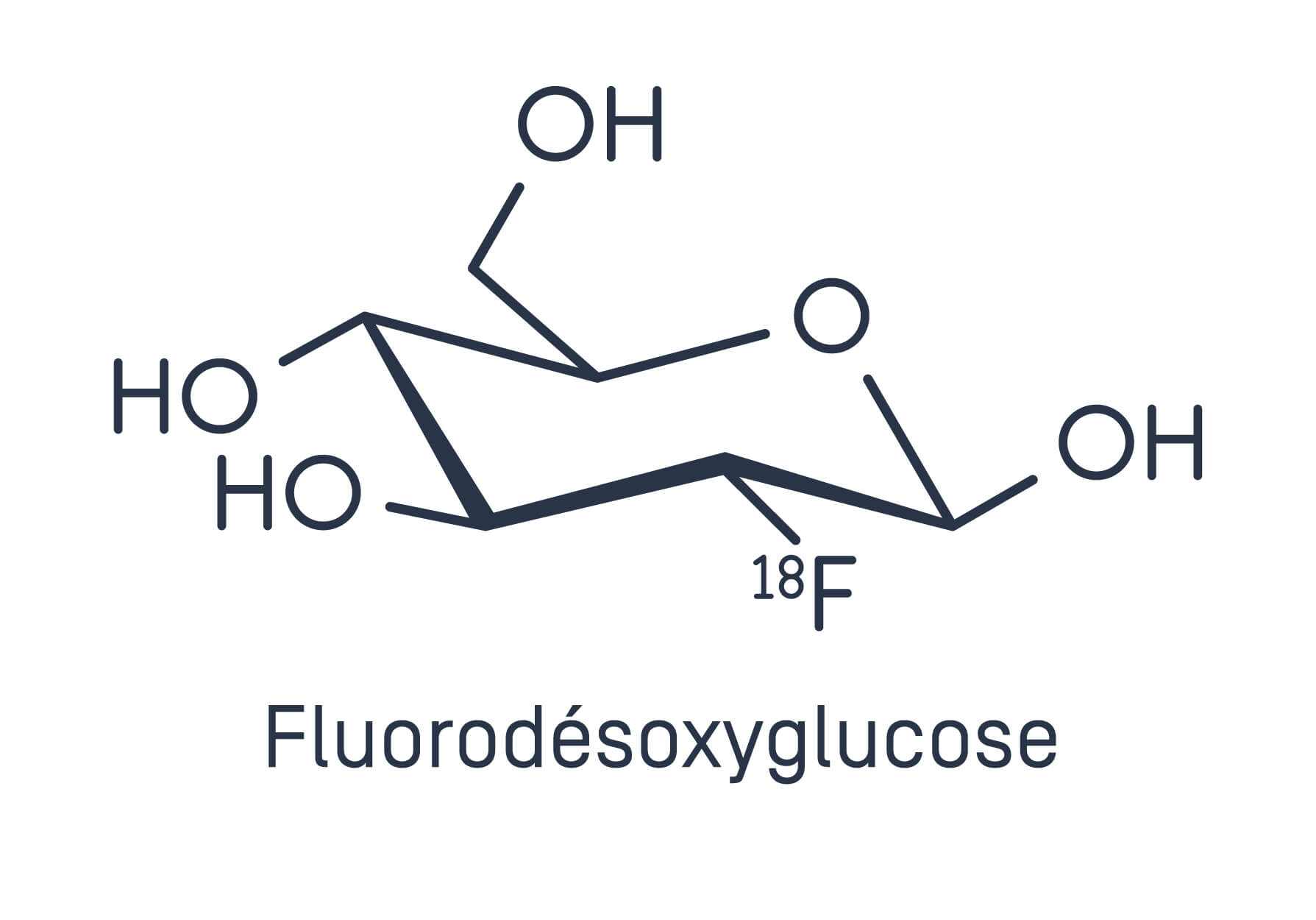
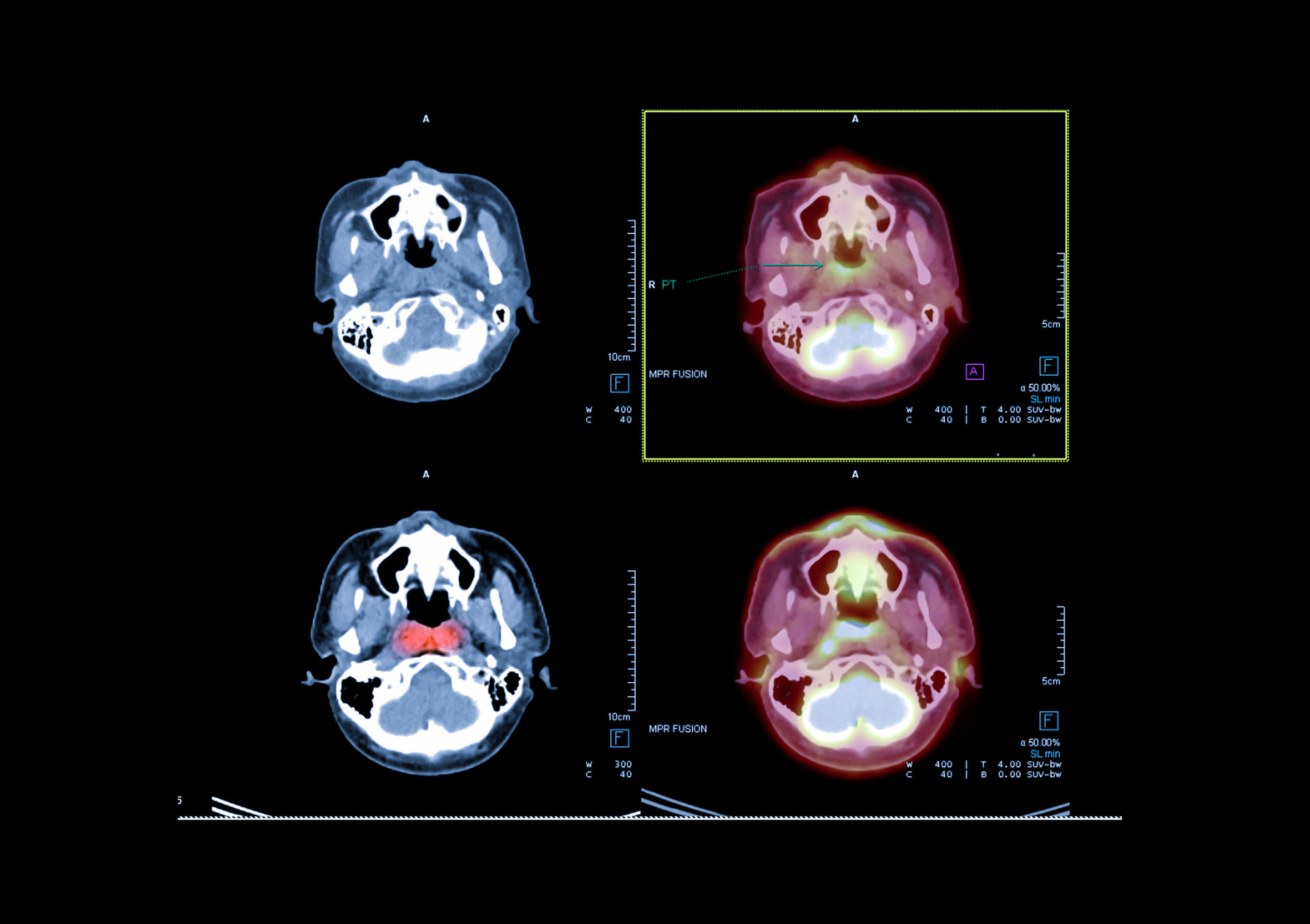
FDG PET-CT is most commonly used in the field of cancer. This is because most cancer cells consume more glucose than normal cells, making them easy to detect on PET scans. The purpose of PET-CT is to:
Locate a primary tumor and assess its aggressiveness.
Perform a staging assessment, i.e., look for possible distant metastases.
Assess the effectiveness of treatment (chemotherapy, radiotherapy).
Detect recurrence in the event of suggestive clinical or biological signs.
The tumors most frequently evaluated by PET-CT are lung, breast, brain, colon, esophageal, and pancreatic cancers, lymphomas, and ENT tumors.


In cardiology and neurology: other applications

PET-CT is also used in cardiology to detect areas of the heart muscle with poor blood supply: it can highlight areas of ischemia or necrosis in the heart muscle. It also evaluates heart function and blood flow in the coronary arteries that supply the heart muscle, thereby helping to determine whether revascularization (bypass or angioplasty) is necessary.
In neurology, PET-CT can reveal abnormalities in the distribution of the radiotracer in the brain in the early stages of degenerative neurological disorders such as Alzheimer's disease. It is also useful in certain cases of epilepsy or brain tumors.


What PET-CT cannot always detect

Certain types of tumors, which are metabolically inactive or small in size, are more difficult to detect using FDG PET-CT. In addition, inflammatory or infectious lesions can capture FDG, making it difficult to differentiate them from tumor lesions.
 
The interpretation of PET images is reserved for specialists in nuclear medicine, who will interpret this data in its clinical and biological context, taking into account all the information at their disposal.

Conclusion

PET-CT is a medical imaging tool that enables more reliable diagnoses, better localization of lesions, monitoring of disease progression, and more relevant treatment guidance. It now plays a central role in oncology, but is also used in neurology, cardiology, and other clinical fields. Well-tolerated, painless, and increasingly accessible, PET-CT is establishing itself as a pillar of precision medicine, helping to understand, monitor, and treat diseases more effectively.
Like any test, it has specific indications and its results must be interpreted by experienced professionals

Questions?

If you have any questions, please do not hesitate to contact us by phone +41 (0)22 719 77 40 or email: secretariat.medecine-nucleaire@latour.ch

FAQ about PET-CT

Can I go home after a PET-CT scan?

Yes. The test does not require hospitalization. You can resume your normal activities, but you should avoid close and prolonged contact with pregnant women and young children (aged 4 and under) for a few hours after the exam, depending on the radiotracer used (12 hours for FDG).

Can PET-CT replace an MRI?

Each imaging exam prescribed provides different information. In some cases, an MRI may provide additional information about your health. This is why you may be prescribed an MRI in addition to a PET-CT scan: one does not replace the other.

Who should I see about these symptoms?

We recommend that you see the following health professional(s) :

Specialties involved