Dr. Gemayel, can you explain what this procedure involves?
The procedure we performed is designed to treat certain aneurysms—that is, dilations of the aorta. In most cases, the treatment is fairly simple: it involves placing a vascular stent graft so that blood flows through this new pathway rather than through the aneurysm. However, if the aneurysm is located in the arteries supplying the kidneys or the intestines, the situation is more complex, as we must ensure that these organs continue to receive adequate blood flow. Otherwise, there is a risk of cutting off blood flow to the affected organ, which can, of course, be fatal.
The solution we’re discussing involves a very specific, custom-made stent that, depending on the case, includes openings (called “windows”), branches, or a combination of both. These components are positioned opposite the target arteries that supply the kidneys, intestines, liver, and spleen, in order to neutralize the aneurysm while preserving blood flow.
The procedure is still rarely performed—is it a recent development?
No, it has been performed for years, but there have been many advancements. The procedure is becoming more widely available, but the facilities that offer it remain, in a sense, an exclusive group, consisting mainly of centers of excellence and major university hospitals.
Initially, this procedure was reserved for high-risk patients, but numerous studies have now demonstrated its effectiveness and long-term results, prompting hospitals to offer it to moderate-risk patients who prefer this technique—which is less invasive and carries fewer risks than traditional surgery. It’s important to remember that we’re primarily dealing with elderly patients who already have lung or heart problems and have undergone other surgeries.
At Hôpital de La Tour, we began performing this procedure in 2017, and we’ve performed several since then. It’s now a treatment we actively promote, especially since it may be the only option for patients who cannot undergo open abdominal surgery. In these cases, we either say, “We’ll do nothing,” knowing that sooner or later the aneurysm will rupture, or we use this technology to treat it. So it has become something we naturally consider.
Why are we currently the only ones in Geneva offering this procedure?
Conventional endovascular surgery is now part of every vascular surgeon’s training, but “windows” are something extra. As for me, I got a head start because I recognized its potential very early on, right from my training. I sought out world-renowned professors in Lille, Paris, London, and Munich to train with them, because I sensed that this was the future of aortic vascular surgery.
As for why Hôpital de La Tour and not the others, I’d say it comes down to infrastructure and expertise. Here, we have an intensive care unit that’s accustomed to caring for cardiac patients with similar comorbidities, and everyone—from the nursing staff to the anesthesiologists—has experience with this type of patient. We also have radiology suites that allow us to perform this type of surgery under optimal conditions—whether in terms of image quality and intraoperative image fusion technology or for verifying the results at the end of the procedure using cone-beam computed tomography. Let’s not forget our interventional vascular radiology department, which is highly effective and at the cutting edge of technology.
In short, we have everything needed to ensure the surgery and follow-up care are conducted under the best possible conditions. Without this range of tools and expertise, a procedure like this would not be possible. We’re truly fortunate here to have every link in the chain working together to ensure the success of these procedures. Conversely, certain decisions or hesitations may also explain why it isn’t yet being performed elsewhere.
So is there still a debate about this technology?
No, I’d say that among those who know how to do it, there’s no longer any doubt. All over the world, when you attend a vascular conference, the majority of sessions are devoted to this. But it’s an advanced technology; it takes a little time for everyone to embrace it and then acquire the skills to perform it routinely. Even in major centers, specialists haven’t necessarily adopted the method yet.
What is the profile of the patients involved?
As I mentioned, these are generally older patients. Most often, these aneurysms occur after age 70. Patients have associated comorbidities, such as coronary or pulmonary problems, and they are often former smokers or people with high blood pressure or high cholesterol.
How is the procedure performed?
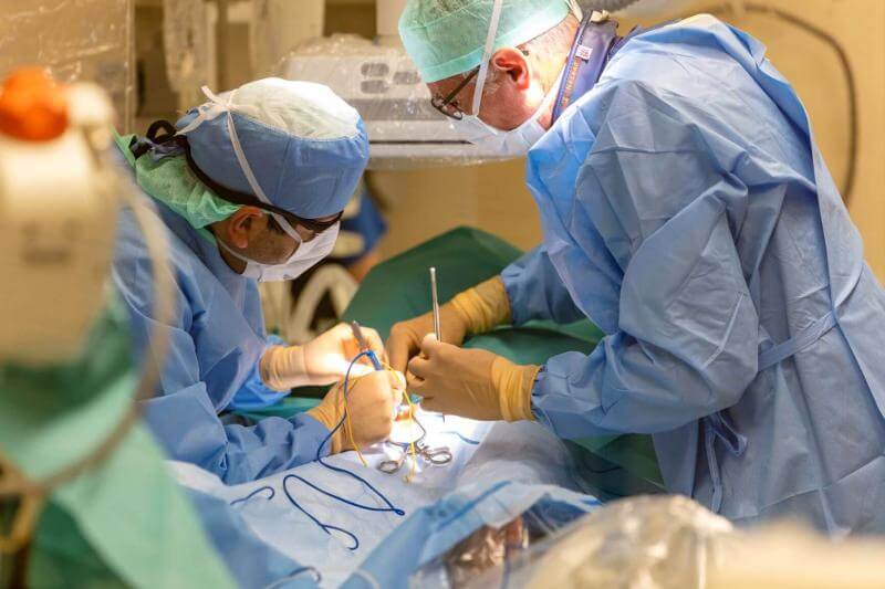
First, based on the CT scan findings, a custom-made stent is fabricated to match the patient’s anatomy exactly. It takes six weeks to two months to receive it. The surgery is then performed under general anesthesia and generally lasts three to four hours, depending on the case. We make a small incision in the groin, near the femoral artery, and proceed upward using guidewires and catheters. In straightforward cases, the arteries run more or less perpendicular to the aorta, and there are no complications; however, in other cases, they run downward or are tortuous, which can prolong the catheterization of these vessels and result in a longer surgery. The prosthesis is positioned—a highly meticulous procedure requiring millimeter-level precision—and then, through the access sites, the various vessels are connected to the prosthesis using stents.
After the procedure, the patient is transferred to the intensive care unit (ICU) for monitoring to ensure there are no immediate complications, particularly involving the stents or access sites. The stay in the ICU is fairly short, which is another advantage of this technique compared to conventional surgery. The patient is then transferred to a regular ward for a hospital stay that is also shorter than usual.
In the case of our most recent patient, the procedure went very well, and he woke up feeling fine right away. He began moving around after 6 hours and was able to return home after 48 hours—which is impressive given his comorbidities and would not have been possible with conventional surgery.
Can there be any contraindications to the surgery?
Yes, mainly anatomical contraindications. There are strict guidelines to follow, the most important of which concern the angles. For example, if the angle between the aorta and the aneurysm is too acute, implanting the prosthesis is technically impossible. Generally, the angle must be no greater than 45°. There is also the issue of access. If the iliac arteries—through which the prosthesis must pass to reach its attachment point—are too small, this poses a problem. This does not always preclude treatment, but it can complicate it by requiring the creation of a surgical tunnel. And then there’s the size of the target arteries. If they’re too small—that is, with a diameter of less than three millimeters—we know that the stent we’re going to place will become blocked and that we won’t be able to save the artery.
That said, when multiple arteries supply an organ, we may, depending on the case, sacrifice one of them. For example, the patient we’re discussing today had three arteries on the left side leading to his kidney. We were able to save two of them, but the third—which was less than 3 mm in diameter—was sacrificed, with no adverse consequences. However, sometimes all the arteries are too narrow, and in that case, it’s a clear contraindication.
What is the postoperative follow-up?
It’s important to remember that when a stent is placed in an aneurysm, it doesn’t disappear. It’s a bit like a defused bomb. To ensure that everything is working properly, long-term monitoring is necessary. An initial CT scan is performed one month after the procedure and is then repeated annually. An MRI or ultrasound can also be used, but a CT scan remains the most sensitive test.
If signs of a problem emerge, intervention is necessary before any damage occurs. For example, if a stent begins to migrate, it may need to be reinforced with another stent. If the aorta or artery dilates, an extension may need to be added using another stent graft. The reintervention rate is 5 to 10 percent, but the vast majority of these procedures will be performed endovascularly. This is a form of maintenance that must be carried out over many years and requires that all patients be monitored regularly.
Comments by Dr. Gino Gemayel, as recorded by Yannick Richter, communications officer.